Parts of the Foot-Anatomy, Functions & Key Details
Introduction to the Parts of the Foot
The parts of the foot play a crucial role in supporting body weight, enabling movement, and maintaining balance. Understanding foot anatomy is essential for anyone dealing with foot parts sole pain, injuries, or general foot care. The foot is a complex structure consisting of bones, muscles, ligaments, tendons, skin, and joints. Proper knowledge of foot anatomy bottom can help prevent injuries and improve overall mobility.
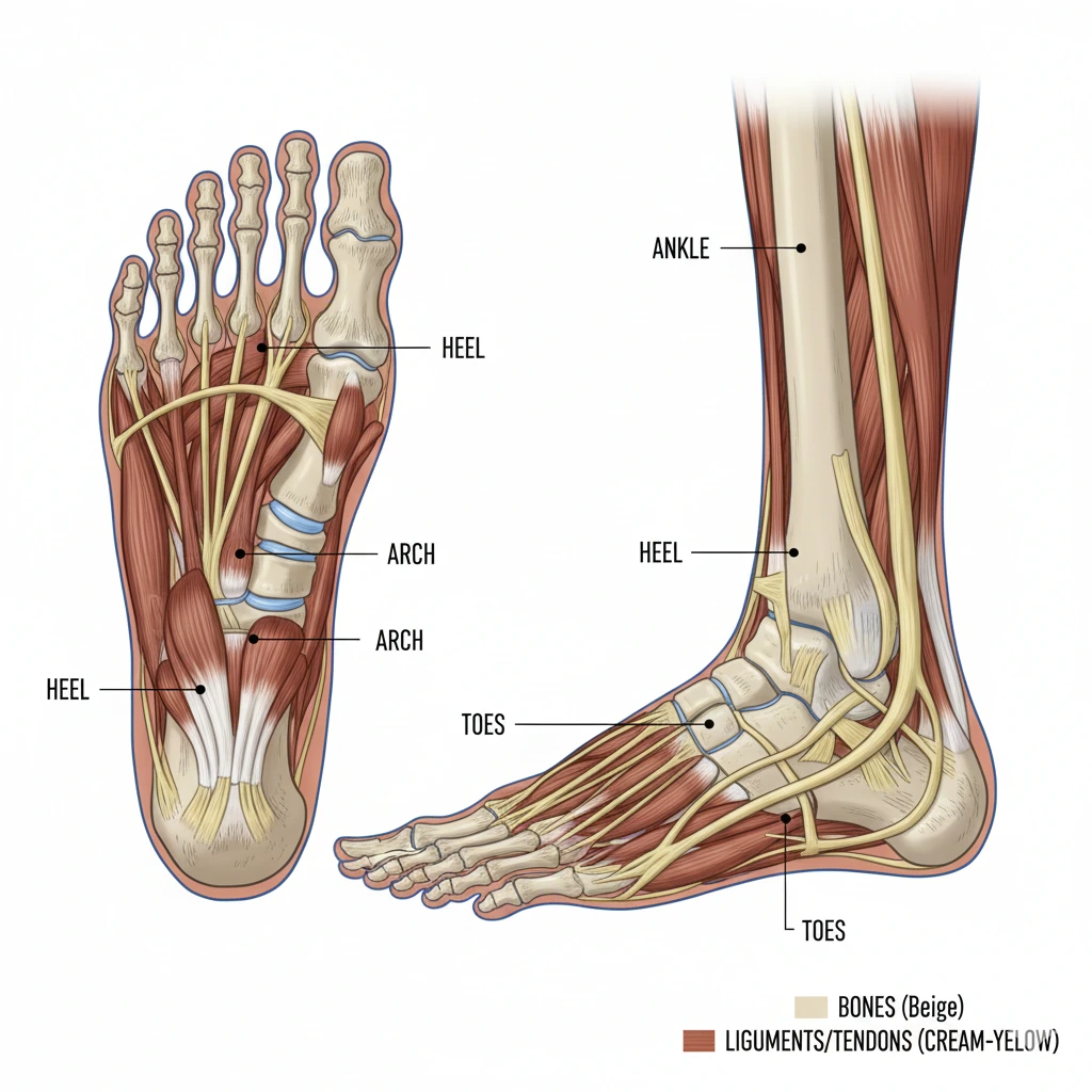
The foot is divided into three main regions: the forefoot, midfoot, and hindfoot. Each region has distinct under foot parts name that contribute to specific functions. The forefoot includes the toes and metatarsals, which assist in propulsion during walking and running. The midfoot contains the arches that distribute weight and absorb shock. The hindfoot includes the heel and ankle bones, providing stability and balance.
Why Understanding Foot Anatomy is Important
-
Helps identify causes of parts of foot sole pain
-
Improves footwear choices for comfort and support
-
Assists in recognizing abnormalities in parts of the foot labeled
-
Essential for athletes and active individuals
Comparison With Other Body Parts
| Body Part | Function | Complexity |
|---|---|---|
| Foot | Support, movement, balance | High |
| Hand | Grasping, manipulation | High |
| Knee | Flexion, support | Moderate |
| Hip | Weight support, mobility | High |
Pros and Cons of Knowing Foot Anatomy
Pros:
-
Reduces risk of injury
-
Enhances posture and gait
-
Helps in early detection of foot disorders
Cons:
-
Complexity may overwhelm beginners
-
Misdiagnosis possible without proper guidance
Customer Highlight
A physical therapy patient in New York said, “Learning the parts of the foot labeled helped me manage my plantar fasciitis and choose the right shoes for long hours of standing.”
Understanding parts of the foot diagram is the foundation for learning the bones, muscles, ligaments, and other essential structures.
Bones of the Foot
The bones of the foot are the structural foundation. Foot bones allow movement, support, and weight distribution. They are divided into three regions: hindfoot, midfoot, and forefoot. Knowing foot parts name with picture is helpful for medical students, fitness enthusiasts, and anyone managing foot parts sole pain.
Hindfoot Bones
-
Talus: connects foot to leg
-
Calcaneus: heel bone supporting weight
Midfoot Bones
-
Navicular, cuboid, cuneiforms
-
Form arches for shock absorption
Forefoot Bones
-
Metatarsals: five long bones connecting midfoot to toes
-
Phalanges: toe bones (proximal, middle, distal)
Foot Bones Table
| Bone | Location | Function |
|---|---|---|
| Talus | Ankle | Connects leg and foot |
| Calcaneus | Heel | Shock absorption, support |
| Metatarsals | Midfoot | Balance, walking |
| Phalanges | Toes | Push-off, stability |
Pros and Cons of Knowing Foot Bones
Pros:
-
Helps identify fractures and injuries
-
Improves exercise and therapy plans
Cons:
-
May be confusing due to multiple small bones
Customer Highlight: A podiatrist in California stated, “Using parts of the foot diagram helps patients understand their conditions better.”
Muscles of the Foot
Muscles control movement and provide stability. Foot muscles are categorized as intrinsic (inside the foot) and extrinsic (from the leg). Understanding parts of the foot and ankle muscles helps manage conditions like plantar fasciitis or toe deformities.
Intrinsic Muscles
-
Flexor and extensor muscles for toes
-
Abductor and adductor muscles for toe spreading
-
Support arches and aid balance
Extrinsic Muscles
-
Tibialis anterior and posterior
-
Peroneal muscles
-
Gastrocnemius and soleus connecting via Achilles tendon
Comparison Table – Foot Muscle Function
| Muscle Type | Location | Function |
|---|---|---|
| Intrinsic | Within foot | Toe movements, arch support |
| Extrinsic | Lower leg | Foot motion, stability |
Pros and Cons of Muscle Knowledge
Pros:
-
Helps in strengthening exercises
-
Reduces parts of foot sole pain
Cons:
-
Detailed knowledge requires study and diagrams
Customer Highlight: “Learning foot anatomy bottom muscles helped me recover faster from a toe sprain,” said a patient in Texas.
Ligaments and Tendons
Ligaments and tendons maintain stability and transmit forces. They connect bones and muscles. Parts of the foot skin overlay these structures, providing protection and sensation.
Important Ligaments
-
Plantar fascia: supports longitudinal arch
-
Collateral ligaments: support toe joints
Key Tendons
-
Achilles tendon: connects calf to heel
-
Tibialis posterior tendon: supports arch
-
Peroneal tendons: lateral stability
Table: Ligaments vs Tendons
| Structure | Function | Common Issues |
|---|---|---|
| Ligaments | Bone to bone | Sprains |
| Tendons | Muscle to bone | Tendinitis |
Pros and Cons
Pros:
-
Essential for stability
-
Prevent overuse injuries
Cons:
-
Injuries can cause prolonged pain
Joints of the Foot
Joints allow flexibility and movement. Main joints include ankle, subtalar, metatarsophalangeal, and interphalangeal joints. Knowledge of foot parts labeled joints helps in managing arthritis and joint pain.
Major Joint Functions
-
Ankle: dorsiflexion, plantarflexion
-
Subtalar: inversion, eversion
-
MTP and IP joints: toe flexibility
Table-Joint Comparison
| Joint | Movement | Location |
|---|---|---|
| Ankle | Flexion/extension | Hindfoot |
| Subtalar | Inversion/eversion | Hindfoot |
| MTP | Toe bending | Forefoot |
| IP | Toe straightening | Forefoot |
Arches of the Foot
Arches provide weight distribution and shock absorption. Main arches include medial longitudinal, lateral longitudinal, and transverse arches.
Pros and Cons of Arch Types
-
Flat feet: stable but prone to overpronation
-
High arches: absorb shock but may strain heels
Table: Arch Functions
| Arch | Function | Common Issue |
|---|---|---|
| Medial | Weight support | Flatfoot |
| Lateral | Stability | Underpronation |
| Transverse | Balance | Pressure points |
Common Foot Problems
Foot issues often relate to parts of foot sole pain or structural problems.
-
Plantar fasciitis
-
Bunions
-
Heel spurs
-
Sprains and fractures
Customer Highlight: “Understanding foot parts name with picture helped me prevent recurring heel pain,” said a Chicago resident.
Foot Care Tips
-
Wash and inspect daily
-
Use supportive footwear
-
Stretch and strengthen muscles
-
Seek professional help for persistent pain
Explore our detailed parts of the foot diagram and learn how each structure works to maintain healthy feet. Take care of your feet today for a lifetime of comfort and mobility.
-
What are the main under foot parts name?
The main under foot parts name include the heel (calcaneus), ball of the foot (metatarsal heads), arch (medial, lateral, transverse), toes (phalanges), and ankle bones (talus). These parts work together to support weight, absorb shock, and enable movement. -
How do the arches affect walking and balance?
The foot has three arches: medial longitudinal, lateral longitudinal, and transverse. They distribute weight, absorb impact during walking or running, and provide balance. Flat feet or high arches can affect posture and may cause parts of foot sole pain if unsupported. -
What muscles control toe movement?
Toe movement is controlled by intrinsic foot muscles, like flexors, extensors, abductors, and adductors, located inside the foot. Extrinsic muscles from the lower leg, such as tibialis anterior and peroneals, also contribute. They allow gripping, pushing off, and maintaining stability. -
How do ligaments and tendons support the foot?
Ligaments connect bones and maintain stability, while tendons connect muscles to bones. Key structures include the plantar fascia, Achilles tendon, and collateral ligaments. They help support the arches, control foot motion, and prevent injuries. -
What causes parts of foot sole pain?
Pain under the foot can be caused by plantar fasciitis, heel spurs, overuse, improper footwear, or injuries. Weak muscles or strain on ligaments and tendons can also lead to discomfort in the foot anatomy bottom. -
How can I strengthen my foot anatomy bottom?
Exercises like toe curls, arch lifts, calf raises, and towel scrunches strengthen the foot anatomy bottom. Regular stretching, proper footwear, and balance exercises reduce the risk of parts of foot sole pain. -
Where can I see foot parts name with picture for study?
You can find labeled diagrams in anatomy books, educational websites, and our detailed parts of the foot diagram on hdpiks.com. These visuals show bones, muscles, ligaments, tendons, and arches for better understanding.